
For a newborn, the difficult part of heel sampling is not simply making blood appear. It is obtaining an acceptable specimen while limiting the depth of the wound and avoiding unnecessary repeat procedures. A neonatal heel lancet should be judged against that whole task.
A shallow-incision device uses a blade that sweeps through a defined path, creating a cut with both depth and width. Compared with a narrower, deeper puncture, this approach can improve collection with less penetration. Trials support advantages for particular devices, not a guarantee for every infant or every product.
To understand the difference, start with the sampling action rather than the brand.
From hand-controlled puncture to a controlled shallow incision
There are three useful approaches to compare. They describe changes in design and control, not a universal historical sequence in which one device disappeared when the next arrived.
| Approach | What controls the action? | What changes for heel sampling? |
|---|---|---|
| Hand-held needle or manual lancet | The operator advances the sharp | Depth and movement depend heavily on technique; there is no automatic cutting cycle |
| Automatic puncture-type safety lancet | A mechanism drives a needle tip or blade through a specified stroke | The device takes over the lancing stroke; specified depth and sharps protection still need checking |
| Dedicated automatic heel incision device | A mechanism drives a blade along a defined cutting path | The design controls incision width as well as depth, allowing a shallow, wider cut intended for heel sampling |
Manual puncture: the operator controls the movement
With a hand-held steel needle, the user’s movement determines how the tip enters and leaves the skin. Training matters, but the needle itself does not provide the automated, preset cutting cycle of a dedicated incision device.
This is the limitation automation addresses: less reliance on judging the lancing movement by hand. It does not make correct positioning, patient selection or training unnecessary.
Automatic safety lancets: control of the stroke
An automatic puncture-type lancet places the lancing movement inside a mechanism. Depending on the model, its needle or blade retracts or becomes shielded after activation. Those are meaningful design changes.
But “automatic” answers only part of the question. The device must also have the right intended use, cutting geometry and depth for the infant. A safety lancet designed for an adult fingerstick is not automatically appropriate for a newborn heel.
Heel incision devices: control of the cut’s shape
The next design question is how to obtain a specimen without relying on a deeper puncture. A sweeping blade adds a defined cutting width while limiting the nominal depth.
A heel incision device can itself be a safety lancet. The useful comparison is therefore between puncture-type and sweeping-incision designs, not between “safe” and “unsafe” products.
Why can a shallow, wider cut be preferable to a point puncture?
A point puncture makes a localised opening. A sweeping blade travels across a short path, creating a shallow incision. The width is designed into the device; the healthcare professional does not manually drag a blade across the heel.
That change gives designers another way to obtain blood: they can specify the width of the opening instead of increasing depth alone. The clinical question is whether that geometry produces the required specimen efficiently, with an acceptable experience for the infant.
Also, a puncture-type safety lancet may contain a blade rather than a needle. Having a blade does not, by itself, establish a sweeping action. Buyers should compare the motion, depth, width and labelled sampling site together.
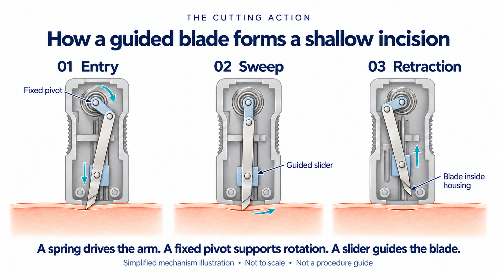
Entry, sweep and retraction
The illustration below separates the cutting cycle into three moments. In this spring-driven design, the arm rotates around a fixed pivot and a slider guides the blade. The blade enters, sweeps through a shallow path and returns inside the housing.
Depth and width are different measurements
A wider incision does not have to be deeper. The selected examples below show why both dimensions matter: a straight blade may have a 2.0 mm depth and 1.5 mm blade width, while a newborn heel-incision configuration has a nominal 1.0 mm depth and 2.5 mm incision width.
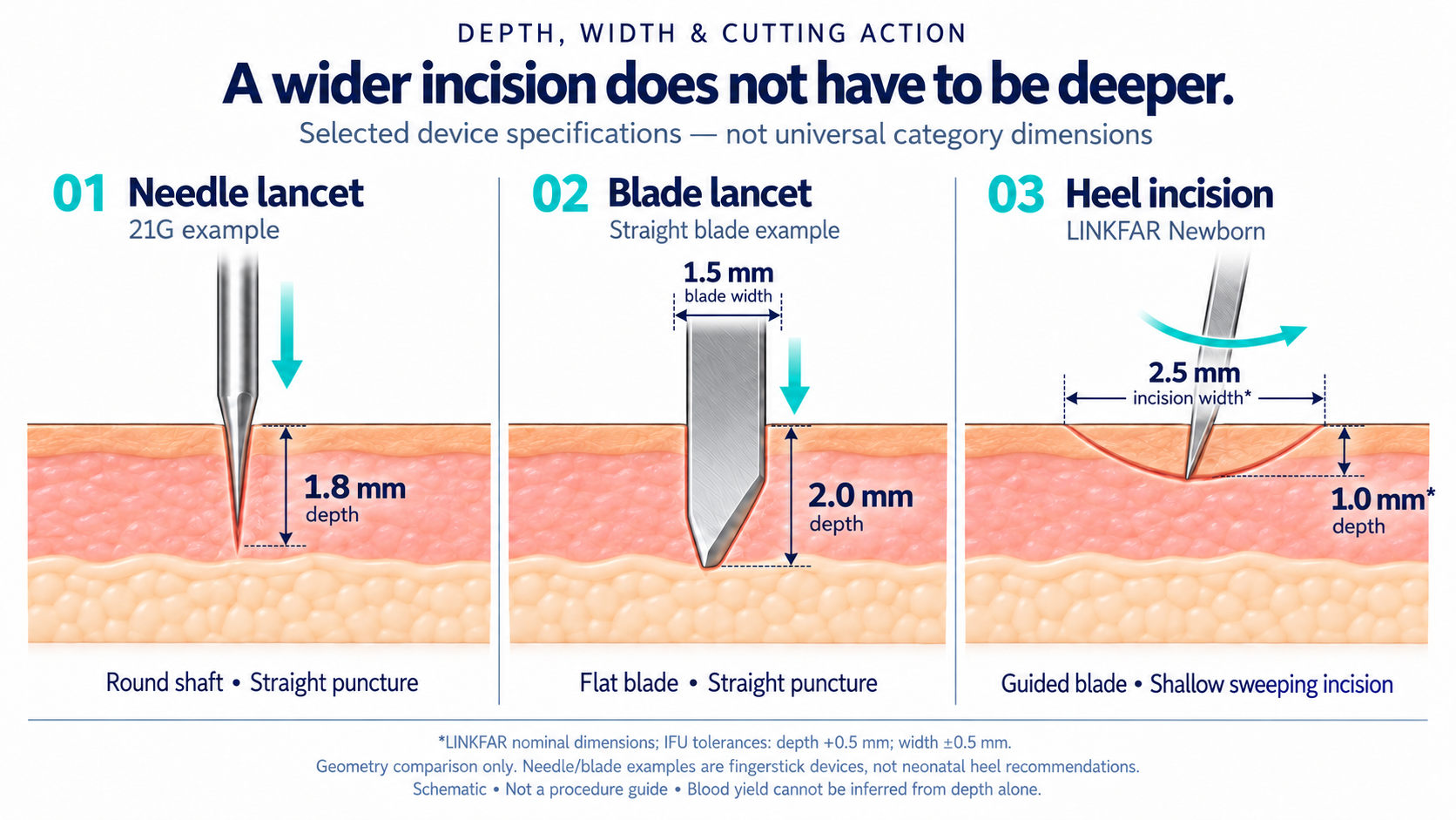
| Illustrated example | Specified depth | Width specification | Sampling-site scope |
|---|---|---|---|
| 21G round-needle lancet | 1.8 mm | No blade-width comparison | Fingerstick example |
| Straight-blade lancet | 2.0 mm | 1.5 mm blade width | Fingerstick example |
| Newborn heel-incision configuration | 1.0 mm nominal | 2.5 mm nominal incision width | Dedicated neonatal heel device |
The needle and straight-blade examples correspond to models 366593 and 366594 in the manufacturer’s specifications and FAQ. The heel-incision values come from the product instructions discussed below, which list a +0.5 mm depth tolerance and a ±0.5 mm width tolerance. Nominal depth is therefore not a guaranteed maximum penetration value.
The comparison explains geometry, not a ranking of blood yield. A 2.0 mm puncture does not inherently provide too little blood, and a 1.0 mm incision does not guarantee enough for every test. Collection results need to be assessed for the device, patient group and specimen requirement.
Depth matters because the heel bone is close
There is limited tissue between the skin and the calcaneus, the heel bone, especially in some premature infants. The clearance is not the same in every baby.
An ultrasound study of 100 preterm and 105 term infants found a skin-to-perichondrium distance below 3 mm in approximately 20% of the preterm group and fewer than 3% of the term group. Among infants born before 33 weeks, the proportion was 38%. These are anatomical observations, not a universal safe-depth calculation. See Arena and colleagues, 2005.
This is why a device intended for neonatal heels deserves more than a “small needle” description. Its depth, selection criteria and interaction with tissue compression matter. A shallow cut addresses the need to limit penetration; it cannot guarantee that bone injury is impossible.
What WHO says about heel-prick depth
WHO’s 2010 phlebotomy guideline states that heel-prick depth should not exceed 2.4 mm and notes a 0.85 mm option for premature neonates. It warns against excessive compression, which can deepen the puncture, and lists calcaneal osteomyelitis among possible complications. See WHO guidance, sections 7.1.2 and 7.1.4.
The 2.4 mm figure is an upper limit in that guideline. It is not a target for every infant, legislation or approval of any particular product.
England’s newborn blood spot programme goes further in its device selection: it specifies an age-appropriate automated incision device, excludes manual lancets and recommends an arched incision. Its illustrations give a 2 mm maximum skin-puncture depth, with a 1 mm maximum device depth for the alternative sites shown for repeatedly punctured heels. These are programme-specific instructions. See the official sampling guidance, section 4.
Device selection must follow the applicable protocol and current instructions. A nominal depth is not automatically a validated maximum projection.
What do clinical studies show about blood flow and repeat sticks?
The strongest reason to consider incision devices is the collection evidence, not how modern the housing looks.
More blood in less time does not automatically mean less pain
In a randomized study of 40 healthy term newborns, an automated incision device yielded more blood in less time and with less haemolysis than a lancet puncture device. The researchers did not find differences in bruising or measured pain responses. See Paes and colleagues, 1993.
That is a useful distinction: collection performance and pain are separate outcomes. Both should be measured.
A shallower, wider incision reduced repeat lancing in one trial
A 2003 randomized trial enrolled 80 healthy term newborns and compared two automatic devices. One made a 1.0 mm deep, 2.5 mm wide incision; the comparator used a 2.2 mm depth and 1.0 mm blade width.
| Outcome | Shallower, wider incision | Deeper, narrower blade lancet |
|---|---|---|
| Sample obtained with one lance | 36/40 infants (90%) | 21/40 infants (53%) |
| Median lancing and collection time | 140 seconds | 215 seconds |
| Median total crying time | 90 seconds | 148 seconds |
Initial-lance pain responses favoured the shallower device; pain during squeezing did not differ significantly. The trial compared QuikHeel with Safety-Flow, was partly manufacturer-funded and did not isolate depth from other design differences. See Shah and colleagues’ study.
These results support evaluating shallow-incision devices. They do not establish that every incision device is better than every puncture lancet, or that every collection will need only one activation. They are category-relevant evidence from named comparators, not results for the product introduced below.
Judge the collection episode, not just the lancet price
The aim is enough suitable blood for the ordered test, not the largest possible blood volume.
A dedicated incision device may cost more than a basic puncture lancet. Its value needs to be assessed over the collection episode: an acceptable sample, collection time, additional devices, laboratory rejection and the infant’s response. A higher unit price is not proof of better performance, but a lower price does not capture the cost of an unsuccessful collection.
For a product evaluation, record the exact device and configuration, first-activation specimen adequacy, collection time, repeat sticks, sample rejection, pain assessed with an appropriate neonatal scale, and adverse events. Handling feedback belongs alongside those measures, not in place of them. Any clinical evaluation needs the appropriate local clinical and ethical oversight.
Device choice also cannot correct every specimen problem. In newborn blood spot screening, poor spots or incomplete information can lead to repeat requests, as the English programme explains. Heel sampling is not the right route for every neonatal test; specimen requirements and the infant’s clinical circumstances still determine the collection method.
Questions buyers ask about heel incision devices
Is a heel incision device different from a safety lancet?
A heel incision device can also be a safety lancet. The useful distinction is the sampling action: a point or blade puncture versus a sweeping incision. Compare cutting path, depth, width, sharps protection and intended neonatal use rather than relying on the product name.
Does “incision” mean cutting deeper?
No. Dedicated heel incision devices can create a wider opening at a shallow depth. Width and depth are separate specifications. Confirm both, including validated limits, against the intended patient group and clinical protocol.
Does a shallow incision guarantee one-stick success or no pain?
No. Trials of particular devices have found collection advantages and, in some outcomes, less pain or crying. Results vary. One-stick success, pain and adverse events require product-specific evidence; a mechanism description alone cannot establish them.
Is WHO’s 2.4 mm limit the recommended depth for every newborn?
No. WHO’s 2010 guidance gives an upper limit, not a target depth, and mentions a 0.85 mm option for premature neonates. Local protocols may be more restrictive. Selection must account for the infant and the device’s current instructions and validated depth limits.
Why consider LINKFAR Heel Blade?
Once a team has decided that a dedicated heel incision device fits its sampling needs, the next step is to assess the actual product.
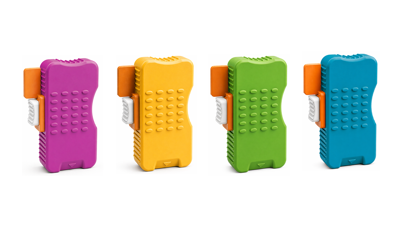
LINKFAR Heel Blade is a single-use neonatal heel incision device intended for healthcare professionals collecting capillary samples from newborns and preterm infants. Its documented design combines a torsion-spring-driven, arc-cutting mechanism with specified nominal incision configurations, a textured housing, a positioning arrow, a removable safety tab and a separate trigger.
Defined incision options
| LINKFAR configuration | Nominal incision depth | Nominal incision width |
|---|---|---|
| Micro-Preemie | 0.65 mm | 1.40 mm |
| Preemie | 0.85 mm | 1.75 mm |
| Newborn | 1.00 mm | 2.50 mm |
These are separate configurations, not user-adjustable depth settings. The range gives buyers defined depth-and-width options to discuss with their clinical team.
The dimensions are nominal, not guaranteed maximum penetration values. The reviewed instructions list a +0.5 mm depth tolerance and a ±0.5 mm width tolerance. Confirm the current controlled specifications, validated maximum projection and applicable instructions before matching a configuration to a clinical depth limit. Configuration names do not replace selection criteria.
For a familiar benchmark, the Preemie and Newborn nominal dimensions match those published for the corresponding BD Quikheel preemie and infant options. That is a dimensional match, not a claim of identical devices or equivalent clinical performance.
Handling features your team can assess
The housing has a textured face and ribbed edges. A raised arrow identifies the centre of the incision. The removable safety tab and separate trigger provide distinct preparation and activation controls.
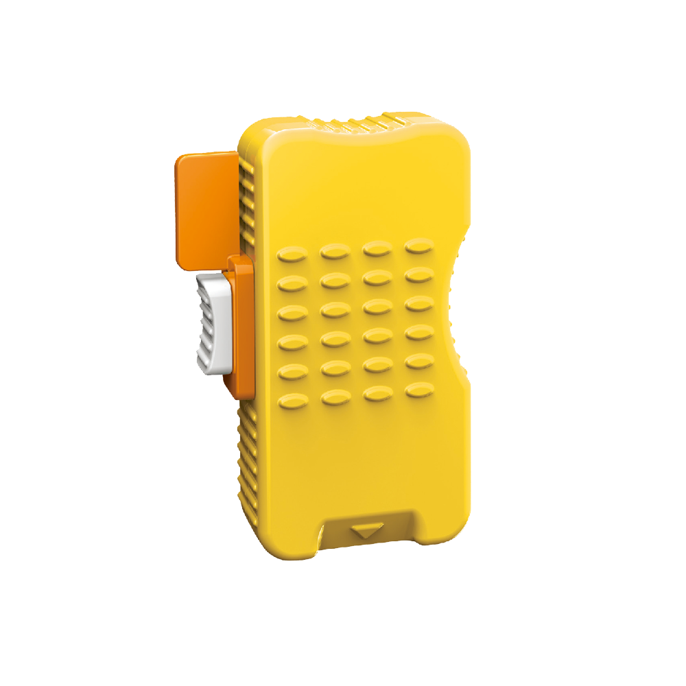
These features give a clinical team concrete things to assess with gloved hands: grip, visibility of the positioning cue and clarity of the controls. The instructions specify single use and deactivation after use. A used device must not be reset or reused.
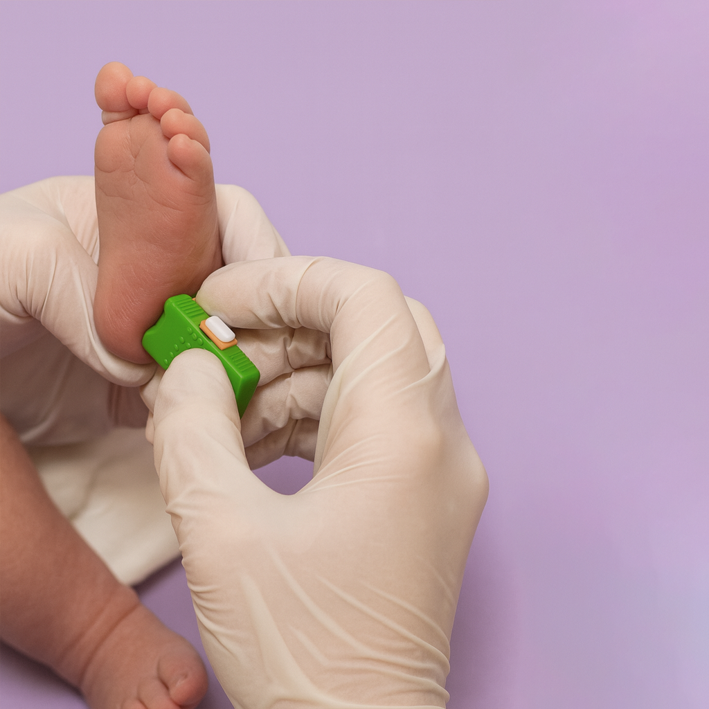
The reason to shortlist LINKFAR is its combination of a documented arc-cutting design, neonatal incision options and identifiable handling features. The clinical studies above explain the value of the incision approach; they should not be read as clinical trials of LINKFAR.
Explore the LINKFAR neonatal heel lancet range or contact LINKFAR with your patient group, destination market and specimen requirements. We can discuss the relevant configuration, current specifications, instructions and samples for your team’s evaluation. For the wider sampling context, see our blood sample collection methods overview.
For healthcare professional and procurement information. This article is not a procedure guide or a substitute for clinical training, local protocols or the supplied instructions for use.
Related guides
-
 Blood Sample Collection Methods: Venous, Capillary and Arterial Blood Collection Explained
Compare venous, capillary and arterial blood collection routes, specimen types, devices and handling considerations for reliable blood sampling.
-
 Dual-spring safety lancets: why PA and PA2 use two springs
The unit price of a safety lancet is easy to compare. The motion of the needle inside it is harder to see, yet that…
-
 LINKFAR upper-arm blood collection enters clinical-stage development
For a patient who needs a small blood sample, the collection appointment can involve more effort than the test itself. Travel, waiting and arranging…
-
 Safety Lancet Manufacturer for Distributors and Retailers: Linkfar Product Guide
If you are a distributor, regional importer, or pharmacy procurement buyer sourcing disposable safety lancets for the Polish market, this page is written for…